Publisher's Synopsis
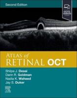
Optical coherence tomography (OCT) is a non-invasive imaging test that uses light waves to take cross-sectional pictures of the retina, the light-sensitive tissue lining the back of the eye (eyeSmart). The technique is recognised worldwide as an essential device for diagnosis, assessment and follow up of retinal diseases and glaucoma. This atlas provides ophthalmologists and trainees with a collection of OCT images to help with the identification, diagnosis and subsequent treatment of common retinal and anterior segment disorders. The images are compiled from the authors' own collections using Plex Elite and Cirrus 6000 technology. Fundus angiography images assist with the understanding of related pathologies. Divided into two sections, the book begins with images illustrating the normal fundus, then numerous different retinal disorders including diabetic retinopathy, macular disorders, retinal detachment, uveitis and toxicities. Section two covers anterior segment disorders, beginning with images of the normal cornea, then illustrating a range of disorders including corneal dystrophies, ocular surface disorders, keratoconus, glaucoma, and trauma. Each section features a multitude of images, each with brief descriptive text.


