Publisher's Synopsis
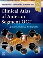
This atlas examines developments in clinical en face imaging, comparing methods and devices and evaluating the most clinically efficient techniques. Divided into three sections, the first part introduces the principles of OCT (optical coherence tomography) and the anatomy and histology of the retina and surrounding area. The second section discusses en face OCT in diagnosing and treating different ocular diseases and disorders. More than 1000 pathological images obtained using different OCT devices are included. The final part describes future developments in the technological and scientific aspects of OCT and their clinical applications. Key points Evaluates clinical en face OCT techniques for numerous ocular diseases and disorders Each case includes pathological images from different devices for comparison Internationally-recognised European and US author and editor team.

